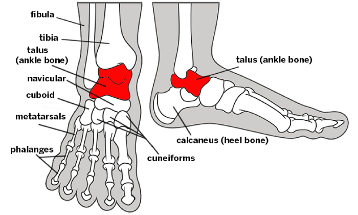
An osteochondral lesion of the talus (OLT) is an area of abnormal, damaged cartilage and bone on the top of the talus bone (the lower bone of the ankle joint). This condition is also known as either osteochondritis dissecans (OCD) of the talus or as a talar osteochondral lesion (OCL). It is often associated with a traumatic injury, such as a severe ankle sprain. However, it can also occur from chronic overload due to malalignment or instability of the ankle joint.
OCLs most commonly occur in two areas of the talus
1. The inside and top part of the lower bone of the ankle (the medial talar dome).
2. The outside and top part of the lower bone of the ankle (the antero-lateral talar dome).
Treatment
Non-Operative Treatment
Non-operative treatment can be successful for non-displaced talar OLTs, especially if the condition is recognized and treated early, and the lesion is relatively small. Younger patients, particularly growing children or adolescents, have a much better chance of healing an OLT compared to adults. There are several non-operative management options for the treatment of osteochondral lesions, including:
- Cast immobilization: If the OLT occurs following an acute injury, initial immobilization in a cast for 4-6 weeks can help reduce stress on the OLT and allow healing. This treatment approach can be initially attempted in non displaced OLTs.
- Physical therapy: working on strengthening the muscles around the ankle, range of motion of the ankle, and balancing (proprioception)
- Protective braces (ex. Ankle Lacer) to decrease stress can also be utilized.
Operative Treatment
- Arthroscopic debridement (cleaning out) and microfracture of the talar OLT. This is the standard operative treatment and leads to good or excellent results in 75-80% of patients with typical talar OCLs (less than 15mm²)
- Osteochondral Autologous Autograft Transfer (OATs Procedure) An OATs-type procedure is reserved for patients who have been treated with arthroscopic cleaning out (debridement) and microfracture and are still not doing well, or patients that have a very large (>20mm²) talar OLT. This procedure may also be called a mosaicplasty. The theoretical advantage of this procedure is that it replaces the damaged cartilage with cartilage and bone harvested from the patient (autograft). The graft is usually harvested from the patient’s knee on the same leg, from an area of that joint that does not bear any load. Patients can develop knee
problems after this procedure. The main disadvantage of this procedure is the prolonged recovery time and increased complication rate, compared to arthroscopic debridement. - Osteochondral Allograft Transfer (i.e., Cadaver): A bone and cartilage plug may also be obtained from a cadaver and transplanted into the OLT. This prevents the need from harvesting bone and cartilage from another part of the body (ex. knee). Osteochondral allografts (Cadaver grafts) have been used to treat large talar lesions with some success. However, the larger the graft, the more likely it seems that it will collapse as a new blood supply is established into the graft after transplantation.
- Autologous chondrocyte transplantation (ACI): There has been an attempt to harvest a patient’s own healthy cartilage, grow the cells in a lab, and then re-implant these cells back into the area where the cartilage has been lost. Unfortunately, this approach in the ankle has not yet met with the type of clinical success that had been hoped for, and is not currently broadly available. Laboratory and clinical work continue in this area.
Our Services
Book Appointment
Book Your Appointment Today
We welcome your questions Do you have questions regarding your own situation? Do you actually want to resolve your problem and not just temporarily cover up the pain?


