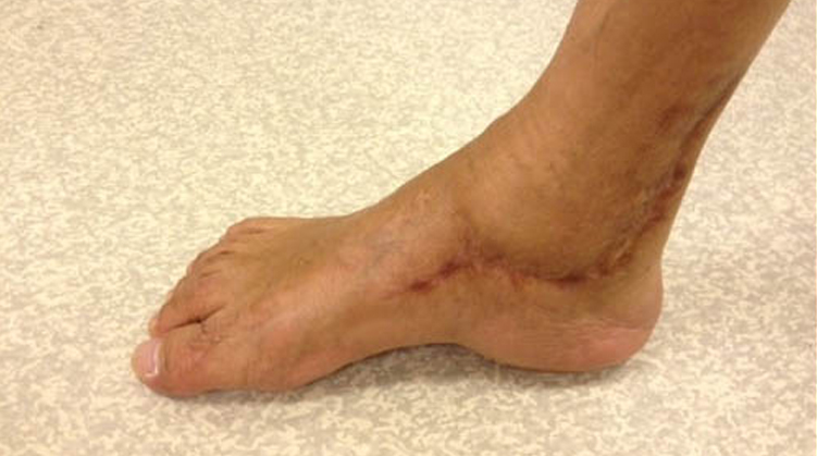
There are many possible pitfalls of clubfoot releases and it is important to recognize the problems and provide proper timely treatment. Late residual deformity following clubfoot releases include: dynamic or stiff supination and forefoot adduction deformities, intoeing gait, overcorrection, rotatory dorsal subluxation of the navicular, vascular insult to the talus with collapse, and dorsal bunion.
The Procedure
The comprehensive surgical release of clubfoot through a Cincinnati incision identifies all tight musculotendinous and ligamentous structures through a wide surgical exposure. Surgical sectioning of these tight structures allows full correction of all the deformities encountered in the clubfoot. The extent and degree of surgical release depends upon the severity of the contractures as well as the lengthening of structures to provide anatomic positioning of the bones of the foot which are then transfixed with pins. Complications from this extensive and complex surgery can occur intraoperatively, immediately postoperatively, or in the longer term.
In our experience, intraoperatively there can be cartilage damage if a scalpel is introduced too deeply into the joint (most commonly the talar head because of its convex configuration), damage to the neurovascular structures, or inadequate positioning of the bones of the foot secondary to incomplete releases or overzealous lengthening. Immediately postoperatively the major complication is vascular or skin problems related to poor circulation from the extensive release or poor positioning of the foot. Later on, there can be substantial scarring resulting in limited range of motion or even loss of part of the foot from vascular or skin loss.
After a period of 6 to 10 weeks of immobilization in a cast, it is necessary to brace the operative foot to prevent the recurrence of the deformity. The brace consists of a hinged ankle-foot orthosis with lateral strap to prevent varus deformity. In addition, postoperatively there can be extensive scarring, stiffness, and loss of motion of the foot which may persist permanently.
A pseudoaneurysm is a rare complication in which a pulsatile mass is encountered either at the level of the incision or on the plantar or dorsal aspect of the foot in association with a rent in one of the major arteries of the foot causing a pulsatile collection of blood. The pseudoaneurysm can work its way to the surface of the skin and cause substantial bleeding from a relatively minor-appearing skin sore. Alternatively, major blood loss can occur in the subcutaneous tissues, which can lead to presentation of children with very low hematocrit. The proper care of the pseudoaneurysm requires recognition of this rare complication and operative treatment.


